The blood-brain barrier (BBB) restricts the passage of many drugs into the brain. This restrictive barrier is created by the presence of many features, such as the tight junctions between the brain capillary endothelial cells and efflux transporter proteins, all of which limit the transport of many compounds into the brain. In the early phase of drug development, cell-based in vitro models are used to predict BBB permeability of new drug candidates.
The MDCK cell line was isolated from Cocker Spaniel kidney tubule. MDCK cells display clear apical-basolateral polarity with well-defined cell junctions, and thus, they are widely used as in vitro epithelial cell models. Two subtypes have been isolated from the parental MDCK strain, MDCKI cells from low passage (~60) and MDCKII cells from high passage (~113). The most striking difference between these two subtypes is that MDCKI monolayer has high trans epithelial electric resistance (TEER), while MDCKII cell monolayer exhibits low TEER. Alkaline phosphatase and γ-glutamyl-transpeptidase activity can be measured in MDCK cells, and since these enzymes are also expressed at the BBB, MDCK cells have been considered to be a useful in vitro model for screen xenobiotics that disrupt the BBB in vivo.
Despite its non-human origin, the MDCK cell line is often the preferred choice in permeability assay because of its shorter culturing time, appropriate prediction of BBB permeability, and presumed closer approximation of passive permeability.
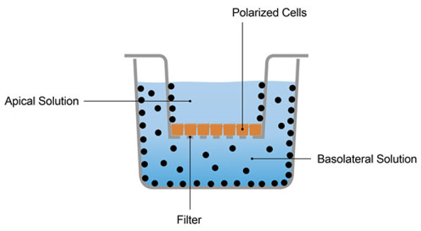 Figure 1. The schematic diagram of MDCK permeability assay.
Use our MDCK permeability assay to identify intestinal or CNS permeability and to investigate drug efflux to find out if your compound is suitable for oral administration or for use as a CNS therapeutic.
MDCK Permeability Assay at Creative Bioarray
Customer provides: Compound identifier and molecular formula;
Cells: MDCK-MDR1 or MDCK-BCRP cells, cultured 4 days to form a monolayer on 24-well PET inserts;
Test compound: Incubation concentration 10 µM, n=2 (flexible);
Format: Unidirectional transport assay and bidirectional transport assay; 24-well plate, volumes used are 0.4 mL apical, and 0.8 mL basolateral, gentle orbital shaking incubator, 2 h at 37ºC;
Controls: Digoxin, quinidine, prazosin, propranolol;
Quantitation: LC-MS/MS;
Readout: Papp (A→B), Papp (B→A), efflux ratio, mass balance;
References
- Chen E. C. et al.; Evaluating the utility of canine Mdr1 knockout Madin-Darby canine kidney I cells in permeability screening and efflux substrate determination. Molecular Pharmaceutics, 2018, 15(11): 5103-5113.
- Volpe D. A. et al.; Drug-permeability and transporter assays in Caco-2 and MDCK cell lines. Future Medicinal Chemistry, 2011, 3(16): 2063-2077.